Rabbit anti-Villin Monoclonal Antibody(039-78)别名宿主反应种属应用分子量免疫原形式浓度纯化方法类型克隆号储存/保存方法存储溶液研究领域使用方法背景说明UniProt
| 概述 | |
| 别名 |
绒毛蛋白;Villin-1; VIL1; VIL
|
| 宿主 |
Rabbit
|
| 反应种属 |
Human, Mouse, Rat
|
| 应用 |
WB:1:1000, IHC:1:2000, ICC:1:250, FC(Intra):1:50
|
| 分子量 |
93kDa
|
| 免疫原 |
Synthetic peptide
|
| 性能 | |
| 形式 |
liquid
|
| 浓度 |
0.5mg/ml
|
| 纯化方法 |
Protein A affinity column
|
| 类型 |
Monoclonal antibody
|
| 克隆号 |
039-78
|
| 储存/保存方法 |
Store at -20℃ for one year.
|
| 存储溶液 |
PBS, 40% Glycerol, 0.05% BSA, 0.03% Proclin 300
|
| 研究领域 |
signal transduction
|
| 使用方法 |
WB: 1:1000, IHC: 1: 2000, ICC: 1:250
|
| 靶标 | |
| 背景说明 |
VIL1 is a gastrointestinal-related cytoskeletal protein that is associated with the microfilament bundles of brush border microvilli. A major structural component of the brush border cytoskeleton, VIL1 binds actin in a calcium-dependent manner. Under normal physiological conditions, villin1 is expressed in epithelial cells of the intestinal mucosa, gall bladder, renal proximal tubules and ductuli efferentes of the testis. Wang et al. report VIL1 to be an epithelial cell-specific anti-apoptotic protein, and to have an
|
| UniProt |
P09327
|
实验结果图

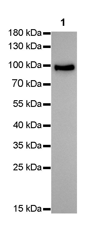
WB result of Villin Rabbit mAb Primary antibody : Villin Rabbit mAb at 1/1000 dilution Lane 1 : HepG2 whole cell lysate 10 µg Secondary antibody: #JP20040 at 1/10000 dilution Predicted MW: 93 kDa Observed MW: 93 kDa Exposure time: 15 seconds

IHC shows positive staining in paraffin-embedded human colon. Anti-Villin antibody was used at 1/2000 dilution, Secondary antibody: #JP20040. Counterstained with hematoxylin. Heat mediated antigen retrieval with Tris/EDTA buffer pH9.0 was performed before commencing with IHC staining protocol.

IHC shows positive staining in paraffin-embedded human kidney. Anti-Villin antibody was used at 1/2000 dilution, Secondary antibody: #JP20040. Counterstained with hematoxylin. Heat mediated antigen retrieval with Tris/EDTA buffer pH9.0 was performed before commencing with IHC staining protocol.

IHC shows positive staining in paraffin-embedded mouse colon. Anti-Villin antibody was used at 1/2000 dilution, Secondary antibody: #JP20040. Counterstained with hematoxylin. Heat mediated antigen retrieval with Tris/EDTA buffer pH9.0 was performed before commencing with IHC staining protocol.

IHC shows negative staining in paraffin-embedded human myocardium(negative tissues). Anti-Villin antibody was used at 1/2000 dilution, Secondary antibody: #JP20040. Counterstained with hematoxylin. Heat mediated antigen retrieval with Tris/EDTA buffer pH9.0 was performed before commencing with IHC staining protocol.

ICC shows cytoplasm staining in HepG2 cells. Anti-Villin antibody was used at 1/250 dilution and incubated overnight at 4°C. Secondary antibody:#JP20025.The cells were fixed with 4% PFA and permeabilized with 0.1% PBS-Triton X-100. Nuclei were countersained with DAPI.

IHC shows positive staining in paraffin-embedded rat kidney. Anti-Villin antibody was used at 1/2000 dilution, Secondary antibody:#JP20040. Counterstained with hematoxylin. Heat mediated antigen retrieval with Tris/EDTA buffer pH9.0 was performed before commencing with IHC staining protocol.

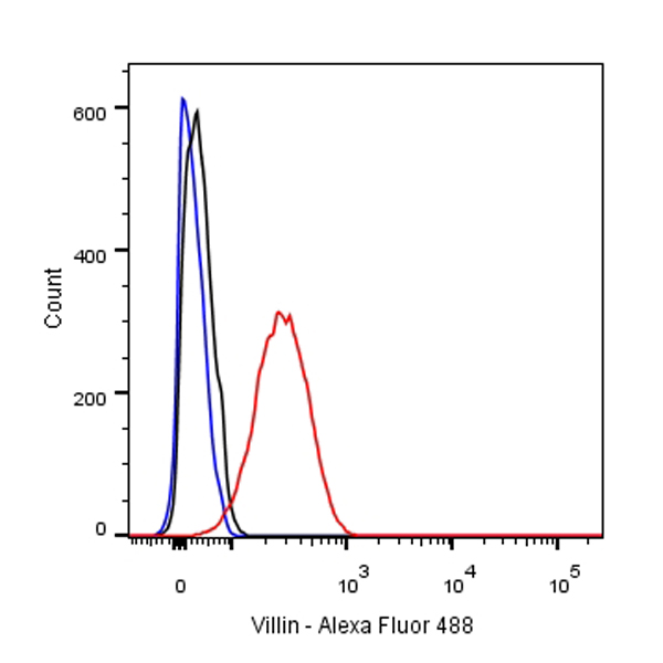
Flow cytometric analysis of HepG2 cells labelling Villin antibody at 1/50 (1ug) dilution/ (red) compared with a Rabbit monoclonal IgG (Black) isotype control and an unlabelled control (cells without incubation with primary antibody and secondary antibody) (Blue). Secondary antibody:#JP20025.
