Mouse anti-PSMA5 Monoclonal Antibody(426CT8.5.1)描述别名宿主特异性反应种属预测反应种属应用分子量类型克隆号同种型储存/保存方法存储溶液研究领域背景说明细胞定位UniProt参考文献
| 概述 | |
| 描述 |
Mouse Monoclonal Antibody (Mab)
|
| 别名 |
PSMA5抗体;Proteasome subunit alpha type-5; Macropain zeta chain; Multicatalytic endopeptidase complex zeta chain; Proteasome zeta chain; PSMA5
|
| 宿主 |
Mouse
|
| 特异性 |
Purified His-tagged PSMA5 protein(Fragment) was used to produced this monoclonal antibody.
|
| 反应种属 |
Human, Mouse, Rat
|
| 预测反应种属 |
B, M
|
| 应用 |
WB~~1:100~500
IHC-P~~1:25 IF~~1:25 |
| 分子量 |
Predicted molecular weight: 26kD
Disclaimer note: The observed molecular weight of the protein may vary from the listed predicted molecular weight due to post translational modifications, post translation cleavages, relative charges, and other experimental factors. |
| 性能 | |
| 类型 |
Monoclonal Antibody
|
| 克隆号 |
426CT8.5.1
|
| 同种型 |
IgG1
|
| 储存/保存方法 |
Maintain refrigerated at 2-8°C for up to 2 weeks. For long term storage store at -20°C in small aliquots to prevent freeze-thaw cycles.
|
| 存储溶液 |
Purified monoclonal antibody supplied in PBS with 0.09% (W/V) sodium azide. This antibody is purified through a protein G column, eluted with high and low pH buffers and neutralized immediately, followed by dialysis against PBS.
|
| 研究领域 |
Cell Biology
|
| 靶标 | |
| 背景说明 |
The proteasome is a multicatalytic proteinase complex which is characterized by its ability to cleave peptides with Arg, Phe, Tyr, Leu, and Glu adjacent to the leaving group at neutral or slightly basic pH. The proteasome has an ATP-dependent proteolytic activity.
|
| 细胞定位 |
Cytoplasm. Nucleus.
|
| UniProt |
P28066
|
| 参考文献 | |
| 参考文献 |
Kottgen, A., et al. Nat. Genet. 42(5):376-384(2010)
Sugiyama, N., et al. Mol. Cell Proteomics 6(6):1103-1109(2007) Olsen, J.V., et al. Cell 127(3):635-648(2006) Beausoleil, S.A., et al. Nat. Biotechnol. 24(10):1285-1292(2006) Hirano, Y., et al. Nature 437(7063):1381-1385(2005) |
实验结果图

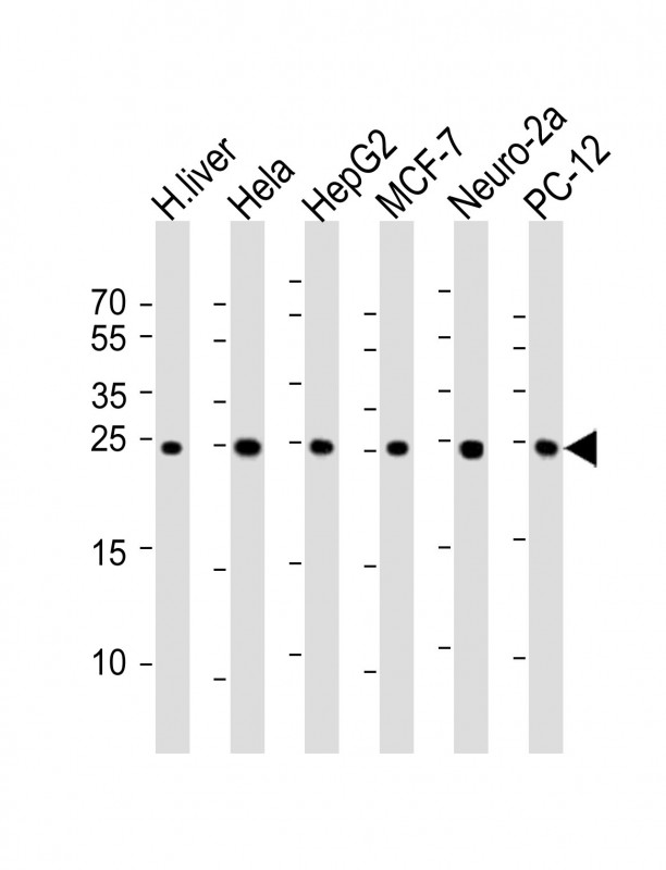
All lanes : Anti-PSMA5 Antibody at 1:1000 dilution Lane 1: human liver lysate Lane 2: Hela whole cell lysate Lane 3: HepG2 whole cell lysate Lane 4: MCF-7 whole cell lysate Lane 5: Neuro-2a whole cell lysate Lane 6: PC-12 whole cell lysate Lysates/proteins at 20 μg per lane. Secondary Goat Anti-mouse IgG, (H+L), Peroxidase conjugated at 1/10000 dilution. Predicted band size : 26 kDa. Blocking/Dilution buffer: 5% NFDM/TBST.

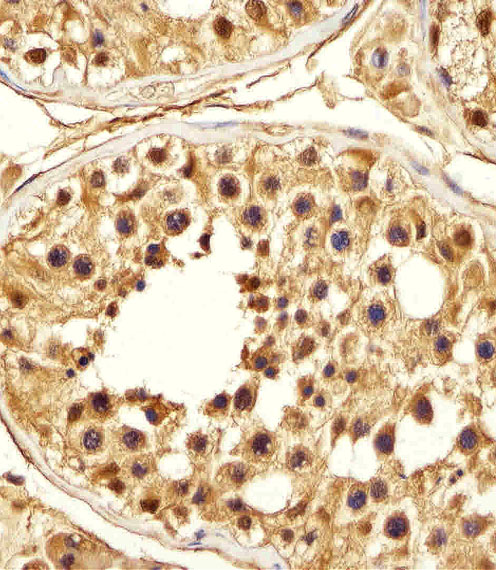
Immunohistochemical analysis of paraffin-embedded H. testis section using PSMA5 Antibody(Cat#JP100217). JP100217 was diluted at 1:25 dilution. A peroxidase-conjugated goat anti-mouse IgG at 1:400 dilution was used as the secondary antibody, followed by DAB staining.

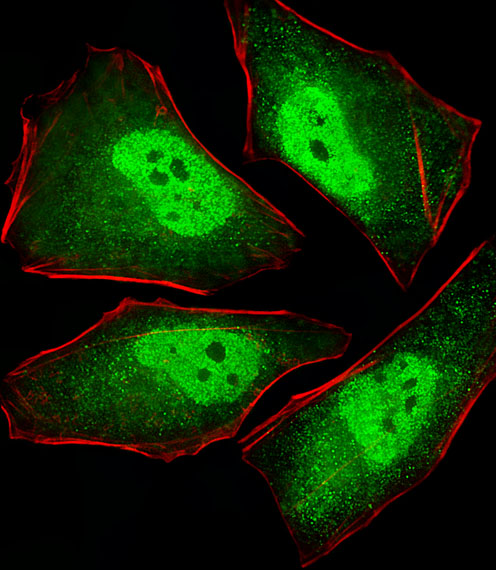
Fluorescent image of Hela cells stained with XAF1 PSMA5 Antibody(Cat#JP100217). JP100217 was diluted at 1:25 dilution. An Alexa Fluor® 488-conjugated goat anti-mouse lgG at 1:400 dilution was used as the secondary antibody (green). Cytoplasmic actin was counterstained with Alexa Fluor® 555 conjugated with Phalloidin (red).

PSMA5 Antibody (Cat. #JP100217) western blot analysis in K562 cell line lysates (35μg/lane).This demonstrates the PSMA5 antibody detected the PSMA5 protein (arrow).
