Mouse anti-RAD51 Monoclonal Antibody(1281CT886.273.179.159)描述别名宿主特异性反应种属应用分子量类型克隆号同种型储存/保存方法研究领域背景说明细胞定位UniProt参考文献
| 概述 | |
| 描述 |
Purified Mouse Monoclonal Antibody (Mab)
|
| 别名 |
RAD51抗体;DNA repair protein RAD51 homolog 1; HsRAD51; hRAD51; RAD51 homolog A; RAD51; RAD51A; RECA
|
| 宿主 |
Mouse
|
| 特异性 |
This RAD51 antibody is generated from a mouse immunized with a recombination protein from human.
|
| 反应种属 |
Human, Mouse, Rat
|
| 应用 |
WB~~1:1000
|
| 分子量 |
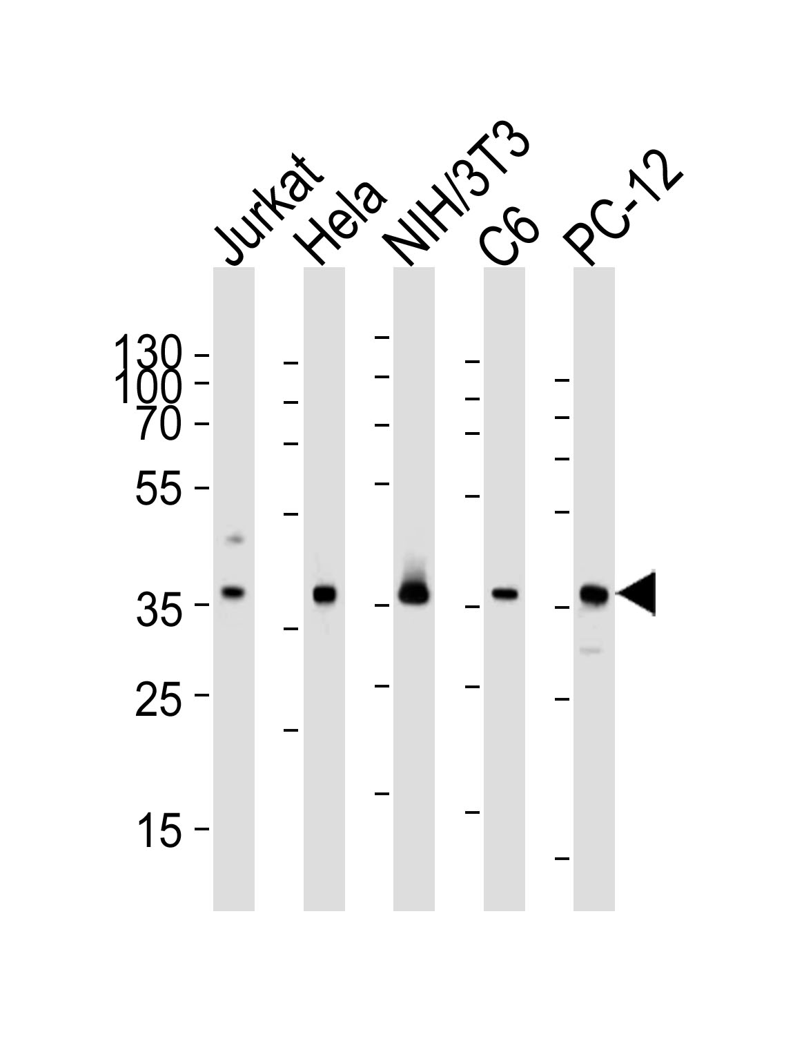
Predicted molecular weight: 37kD
Disclaimer note: The observed molecular weight of the protein may vary from the listed predicted molecular weight due to post translational modifications, post translation cleavages, relative charges, and other experimental factors. |
| 性能 | |
| 类型 |
Monoclonal Antibody
|
| 克隆号 |
1281CT886.273.179.159
|
| 同种型 |
IgG1,κ
|
| 储存/保存方法 |
Maintain refrigerated at 2-8°C for up to 2 weeks. For long time storage store at -20°C in small aliquots to prevent freeze-thaw cycles.
|
| 研究领域 |
Crown Antibodies
|
| 靶标 | |
| 背景说明 |
Participates in a common DNA damage response pathway associated with the activation of homologous recombination and double-strand break repair. Binds to single and double-stranded DNA and exhibits DNA-dependent ATPase activity. Underwinds duplex DNA and forms helical nucleoprotein filaments. Part of a PALB2- scaffolded HR complex containing BRCA2 and RAD51C and which is thought to play a role in DNA repair by HR. Plays a role in regulating mitochondrial DNA copy number under conditions of oxidative stress in the presence of RAD51C and XRCC3.
|
| 细胞定位 |
Nucleus. Cytoplasm. Cytoplasm, perinuclear region. Mitochondrion matrix. Cytoplasm, cytoskeleton, microtubule organizing center, centrosome. Note=Colocalizes with RAD51AP1 and RPA2 to multiple nuclear foci upon induction of DNA damage. DNA damage induces an increase in nuclear levels. Together with FIGNL1, redistributed in discrete nuclear DNA damage-induced foci after ionizing radiation (IR) or camptothecin (CPT) treatment Accumulated at sites of DNA damage in a SPIDR-dependent manner
|
| UniProt |
Q06609
|
| 参考文献 | |
| 参考文献 |
Shinohara A.,et al.Nat. Genet. 4:239-243(1993).
Yoshimura Y.,et al.Nucleic Acids Res. 21:1665-1665(1993). Schmutte C.,et al.Cancer Res. 59:4564-4569(1999). Wang W.W.,et al.Cancer Epidemiol. Biomarkers Prev. 10:955-960(2001). Park J.Y.,et al.Nucleic Acids Res. 36:3226-3234(2008). |
实验结果图

Western blot analysis of lysates from Jurkat, Hela, mouse NIH/3T3, rat C6, PC-12 cell line (from left to right), using RAD51 Antibody(Cat. #JP100449). JP100449 was diluted at 1:1000 at each lane. A goat anti-mouse IgG H&L(HRP) at 1:3000 dilution was used as the secondary antibody. Lysates at 35μg per lane.
